
To quote Malcolm Gladwell, I think I witnessed a “Tipping Point” yesterday. The standard of practice for determining the presence or absence of pneumonia and pneumothorax has always been a chest radiograph. Determination of fluid collections by ultrasonography in the chest or abdomen, the domain of the Pediatric Radiologist.
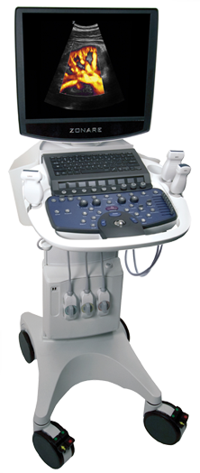
Yesterday however, I was introduced to the use of bedside Point of Care Ultrasound (POC U/S) for the diagnosis of all of these aforementioned conditions. I spent the morning at a course on POC U/S for the Neonatologist and with that became comfortable much sooner than I expected with making such diagnoses based on some very straightforward criteria. Now before you think I am getting ahead of myself, I do not believe for a second that I am now competent to use U/S to replace radiographs as the “gold standard” in my practice. It has led me to consider though how one might go about reaching this level of confidence but I will share more on that later.
An example of how one can use ultrasound to exclude a pneumothorax is shown here https://www.youtube.com/watch?v=ws7DI4ZGQCo. The presence of bilateral “lung sliding” of the parietal and visceral pleura accompanied by “comet trails” excludes a pneumothorax. The absence of these findings is suggestive of a pneumothorax although other pathological findings can be present necessitating a chest x-ray. Think of how many times a patient develops a sudden increase in FiO2 and you do a chest x-ray to exclude a pneumothorax before increasing PEEP. This could change with acquisition of such technology.
The first question to really ask before taking the leap to replace or augment the classic chest radiograph is to look at the literature. With respect to the Neonate, the body of literature is not large but there are a couple of recent papers that are worth mentioning. The first is by Pereda MA et al. Lung Ultrasound for the Diagnosis of Pneumonia in Children: A Meta-analysis. (http://1.usa.gov/1CM24Sv) The review examined 9 studies of which 2 were in the neonatal population. The findings were intriguing. The sensitivity was 96% and specificity 93% when compared to the “Gold Standard” of either CXR or CXR with clinical diagnosis. To put this into context, there were 765 children who would have had thousands of x-rays during their hospital stay. This technology could reduce the number to ionizing radiation exposure by an extraordinary amount given the accuracy of the test compared to standard x-rays. The benefit of such reduction can be immediately appreciated when one considers that children are at an increased risk of malignancy compared to adults from ionizing radiation. Free supporting publication here.
The second paper to highlight is by Lovrenski J. Lung Ultrasonography of pulmonary complications in preterm infants with respiratory distress syndrome. Free supporting publication here
In this paper 120 patients with respiratory distress syndrome had U/S performed in the first 24 hours of life and then follow-up as indicated. Of this sample, 47 had complications including hemorrhage, pneumothorax, pneumonia, atelectasis and bronchopulmonary dysplasia. Of this group 45 were detected by ultrasound with the only two failures being small pneumothoraces. There were 512 ultrasounds performed compared to 612 clinically indicated chest x-rays during the same period. Again the potential reduction in ionizing radiation is astounding!
To be clear I am not suggesting that we do away with the chest x-ray but rather there is a great potential for reducing the need for the urgent chest x-ray when patient status changes. Depending on the situation in the unit you work in, an x-ray from the time it is ordered till the time it is performed and processed can be 15-30 minutes or more. The time it takes to perform a bedside ultrasound exam is 2-3 minutes at most making this modality at least in my mind a great first line option.
As I mentioned earlier though I am not an expert but merely an enthusiastic Neonatologist. This is not meant to replace the Pediatric Radiologist and in fact I would like to stress that I believe it is best suited to questions in which a binary yes/no answer is being asked. Does the patient have a pneumothorax, atelectasis, fluid in the abdomen or chest; all yes/no. Does the patient have a tumour in the liver and if so what type? That is a different kettle of fish and deserving of a Pediatric Radiologist’s opinion.
How do we go from “good to great” and utilize this technology accurately and safely. Clearly we are in need of practice, which can only come through training by qualified people who have demonstrated proficiency in the field. Thankfully in our situation we have access to individuals who have taught courses internationally and are willing to provide the training locally to us. Our strategy will include monthly U/S rounds supervised by these two experts who will provide a small group of us “superusers” with the training and ultimately confidence to put this into routine clinical practice. As confidence rises, the use of POC U/S to obtain vascular access will develop as will performance of such procedures as a bladder tap under direct visualization to improve our success rate. I suspect new uses will develop over time as we are at the forefront of this technology. There are courses offered abroad but like the paucity of Neonatal research in the area there are very few if any such programs for the Neonate specifically.
What I can say for sure is that the benefits of POC U/S are vast and I believe our patients will benefit both during their hospital stay and into adulthood due to the reduction in ionizing radiation that I see coming for these fragile infants. I for one am excited and energized by the future of this field and look forward to an expanding body of literature on the topic.
Hi. My hospital reality is that a chest rx delays even.24 hours. We are a litle hospital from a public health that is quite poor. Ultrasound is limited at the radiologic department. Its ssuch.a.dream when I think in a hospital like yours. Again congrats to you. I.am so happy to read and to know that therae are other people interested in no agresive treatments.
thanks for sharing your knowledge.
There are courses available in many parts of the world. If you want it you can have it but it takes a little effort! Would give immediate results.
It is really a nice share. I have been looking for this to know from long. Thank you for sharing